Whoa.... Our first practical in term 3!
Today, we carried out an experiment, involving the separation technique chromatography. It was stated on the worksheet that there were 3 suspects caught with and each possessed a different brand of black ink- brand A, brand B and brand C. We were told to use chromatography to find out which suspect wrote the secret message.
As it was my first time trying out chromatography, I was bursting with excitement (as usual :P)
When we placed the chromatography paper into ethanol, the different components of the black ink started spreading up the chromatography paper, which was seriously amazing.
Through this practical, I also understood how useful chromatography can be. Firstly, chromatography can separate the components in a sample. Secondly, chromatography csn determine if the sample is pure. Lastly, it can identify the substances present in a sample.
Many scientists use chromatography to detect traces of unpermitted addictives in foodstuffs, etc. Also, the police usually use chromatography to identify poisons/ drugs in urine samples.
Chromatography is really interesting, eh?
1P14: Inside of Cells
Today's practical is so cool! We actually get to use the light microscopes! This practical wanted us to prepare our own samples, and to observe its cell structure under the microscope. The sample Mrs Koh wanted us to prepare was the cheek cells. We just needed a toothpick to scrape out some cheek cells from the inside of our mouths and then stain them with the iodine solution. Much easier than you thought, right?
Anyway, this was how it looked like under the microscope:

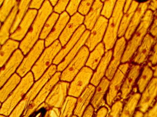
Another sample we made was the onion skin sample. To do so, we must peel of a thin, transparent layer of skin from the oniom using a pair of forceps. Again, we must stain the cells with iodine since its brown.
Peeling the skin was quite difficult as I always peel of a layer which is too thick. Luckily, after a few tries with the help of my partner, I finally managed to do it. This was how it looked like under the microscope:
Observing cells under the microscope was really cool and hopefully there will be more of such practicals :D
1P15: Bouncy Raw Egg with Moving Waters
Today, we did an experiment, mainly to demonstrate osmosis in living cells. To do so, we needed a quail egg and some diluted hydrochloric acid.
Why a quail egg?
The quail's egg is enclosed by a thin egg membrane and is protected by an egg shell. The egg shell is made of calcium carbonate, which is partially permeable.

We had to handle the acid carefully as it is corrosive and I don't think anyone wants their hand getting corroded :P
Anyway, we needed to place the egg in a cup of hydrochloric acid. Then, surprisingly, the egg shell started peeling off by itself! Not only that, bubbles of colorless and odourless with white froth was seen.
Actually, all these happened due to osmosis. Water has moved from a region of higher water concentration in the beaker to a region of lower water concentration in the egg across the partially permeable egg membrane. Thus, it became turgid too.
Mrs Koh wanted us to then place the peeled egg into a beaker of water. Then, after 2 days, we needed to go back again to see the change in the egg's size and hardness.
1P17: Extracting Chlorophyll
Today, we went to the lab to carry out an experiment on extracting chlorophyll from leaves. The best thing is.... I get to use the Bunsen burner again! :D
To extract chlorophyll, we must first heat the leaf in boiling water. This is to kill the leaf and ensure that no more chemical reaction is taking place. Then, we must place the leaf in a beaker of methylated spirits in a 250mL beaker.
Soon, the methylated spirits started to become green as the chlorophyll is now in solution in the methylated spirits. The leaf looks something like that:

To check if our experiment was a success, we tested it with iodine. Starch was present on the green parts of the leaf but not on the yellow/white parts of the leaf.

1P18: Artificial Intestines
Today's practical was the last one for this term :( (Time really flies!)
Anyway, today's experiment was to investigate the reason why food needs to be digested. To do so, we needed a visking tubing and some starch and glucose solution. Firstly, we poured some starch and glucose solution into the visking tubing. Afterwards, we placed it in a boiling tube filled with distilled water. The experiment set up looks something like this:

After around an hour, we tested the boiling tube for any presence of starch and glucose. To test for starch, we added a few drops of iodine solution into the test tube sample; the iodine solution will turn from yellow-brown to blue-black colour if there is a presence of starch.
We also tested the test tube sample for any reducing sugar. We added a few drops of Benedict's solution into a test tube sample, and place the test tube ina hot water bath until just boiling. A brick red precipitate will be formed if there is a presence of reducing sugars.
The iodine did not change colour as the starch was still trapped in the semi-permeable visking tubing. However, when the water was tested with Benedict's solution, it turned into a brick-red precipitate. This is because sugar molecules are small enough to diffuse across the semi-permeable membrane (as in the visking tubing) while the starch molecules are just too large. This shows the importance of digestion in breaking down large complex molecules to smaller diffusible substances that can be absorbed into the bloodstream.
No comments:
Post a Comment